![]()
Ron Dimock's Home Page

Ronald V. Dimock, Jr.
Thurman D. Kitchin Professor of Biology
Department of Biology
Wake Forest University
Winston-Salem, NC 27109
Education:
- BA Zoology, University of New Hampshire
- MS Biology, Florida State University
- PhD Biology, University of California at Santa Barbara
Director,
Wake Forest University Mussel Research Center
Current Research:
- Physiological Ecology and Behavior of Freshwater
Mussels
Recent
Publications from My Lab on Adult and Juvenile Unionid
Mussels
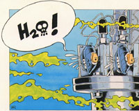
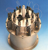
MUSSELS: guardians of your water quality!!
Design by: W. Rebergen,Delta
Consult,Kapelle, The Netherlands. On the right is a
commercial water pollution monitor
based on mussel gaping. (Delta Consult, Kapelle, NL)
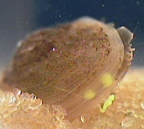
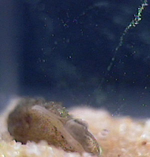
The aquarium facility for housing adult and juvenile mussels in my laboratory:
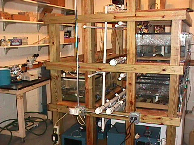
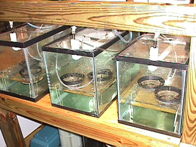
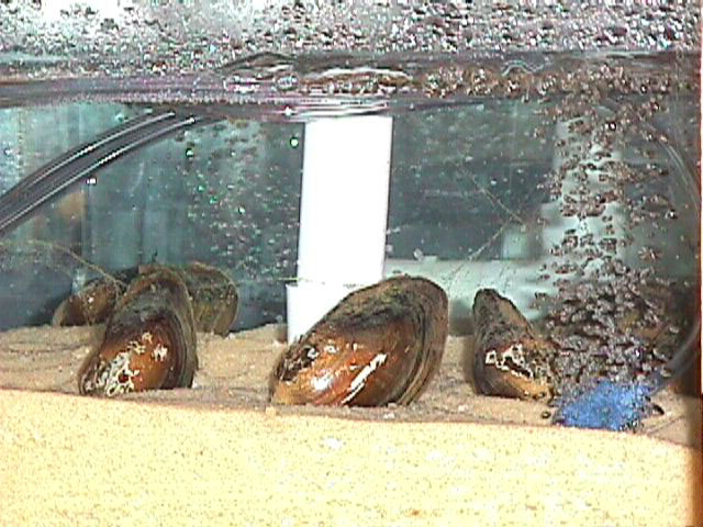
Chambers housing juveniles in a down-welling system (left) and adult U.imbecillis releasing glochidia larvae (visible as mucous strands from exhalant siphons).
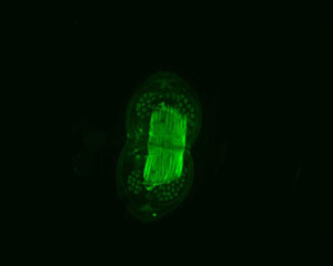
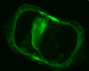
Glochidia larvae of the
mussel Utterbackia imbecillis
with the adductor muscle stained with a fluorochrome
that binds to actin filaments. The valves are fully open. Larvae are about 280
microns in length (axis parallel to the hinge).
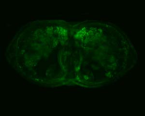
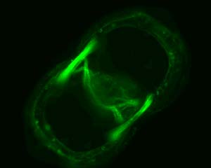
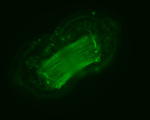
Larva at day-4 of metamorphosis (upper left), with adductor muscle gone. Juvenile (upper right) 7 days post-metamorphosis showing new anterior and posterior adductor muscles. The first 3 pairs of gills filaments are partially visible (especially to the right of the mid-central foot). Lower image is of a juvenile 10 days post-metamorphosis, showing brightly fluorescing adductor muscles, some pedal musculature (center) and the heart within the pericardial sinus (toward 4 o'clock, just right of center of image, near posterior adductor).
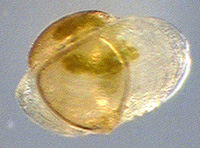
3-week old juvenile Pyganodon cataracta :
anterior to the right, total length about 450 microns; subtriangular
part of shell is original larval shell; brownish-green is silt and algae in
stomach and digestive glands.
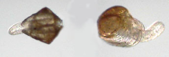
Juvenile P. cataracta : about 5 weeks old. Individual on right is about 700
microns. Note new shell growth flanking foot on animal at right.
10 week old P. cataracta approximately 3.5 mm
long, showing well developed inhalant and exhalant siphons. Animal is in
process of rejecting a mass of yellow latex beads that it has filtered out of
suspension.
Same juvenile as above showing a plume of latex beads in the
flow from the exhalant siphon.
- Physiology and Behavior of Symbiotic Mussel-Mites:
Genus Unionicola
Recent
Publications from My Lab on Unionicolid Mites
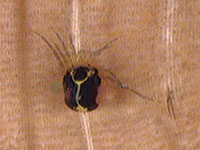
The water mite U. formosa
on the gill of its host mussel P. cataracta
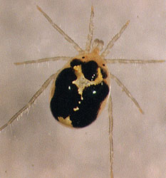
Photo by Ginger Fisher -- Female U. formosa
Web
site devoted to Arachnology which of
course includes the study of the Acari, the
Order of mites, ticks, etc.
Mussel/mollusc related web sites:
Check out UNIO, a Listserver for anyone interested in the biology of freshwater mussels.
The Freshwater Mussel Conservation Society web site is an excellent source of information, with a lot of great links to useful resources on the web.
A nice molluscan resource site developed by Deborah Wills
A terrific gallery of mussel images maintained by Chris Barnhart, Unio Gallery
Home Page of the American Microscopical
Society AMS, a great place for
Invertebrate Biologists to gather.
 Click
on this image if you are interested in Integrative and Comparative Biology
Click
on this image if you are interested in Integrative and Comparative Biology
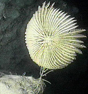

An isocrinid crinoid at a depth of
800 feet near the wreck of the "Kirks Pride", Grand Cayman Island.
The pinnate arms are extended in feeding posture. Current is flowing from right
to left.
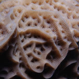
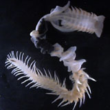
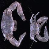
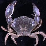
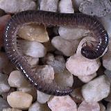
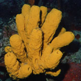
Assorted Invertebrates (Upper left and lower right photos by Craig Nelson)
Courses I Regularly Teach at Wake Forest University:
Home pages of a few of my former MS or PhD students
The
Who and Where of My Former
Graduate Students
Department of Biology Home Page
- Revised November 2011
![]()
[Home] [Directory] [Search] [Visitor] [Computing] [Details] [Comments]